Home
/ Diagram Of Hip.and Back.muscles - Hip Pain Explained Including Structures Anatomy Of The Hip And Pelvis - The hip muscles are going to be slip into hip muscles and gluteal muscles.
Diagram Of Hip.and Back.muscles - Hip Pain Explained Including Structures Anatomy Of The Hip And Pelvis - The hip muscles are going to be slip into hip muscles and gluteal muscles.
Diagram Of Hip.and Back.muscles - Hip Pain Explained Including Structures Anatomy Of The Hip And Pelvis - The hip muscles are going to be slip into hip muscles and gluteal muscles.. It is opposite from the chest, and the vertebral column runs down. In human anatomy, the muscles of the hip joint are those muscles that cause movement in the hip. Muscle anatomy for bodybuilding 12 photos of the muscle anatomy for bodybuilding chest muscles anatomy for bodybuilders, muscle anatomy and bodybuilding, muscle anatomy for bodybuilding, muscle anatomy workout book, muscle anatomy workout pdf, human muscles. Dislocation of the hip joint. Diagram of muscles and anatomy charts.
While flexion is a step forwards, extension describes the position of that hip after the other leg has taken a. The levator ani muscle along with a second muscle forms the pelvic floor. Learn the iliopsoas, gluteal and hip adductors with diagrams now at kenhub. Back muscles anatomy lower back muscles anatomy human anatomy. They begin under the gluteus maximus behind the hip bone and attach to the tibia at the knee.
Hip Anatomy And Function from www.hipkneetumoursurgery.com Learn the iliopsoas, gluteal and hip adductors with diagrams now at kenhub. Common hip and back pain causes include injury to muscles from overuse, disc injury/degeneration, or spinal stenosis. To learn more about the lower back anatomy of the spine, please watch this video. Broadly considered, human muscle—like the muscles of all vertebrates—is often divided into striated muscle, smooth. This is a table of skeletal muscles of the human anatomy. Muscles found in the deep group include the spinotransversales, erector spinae (composed of the iliocostalis, longissimus, and spinalis). Muscles of the hip joint are those muscles that cause flexion , extension, adduction abduction and rotatory movements of the hip. Learn with flashcards, games and more — for free.
The muscles in the forearm and palm thenar muscles all work together to keep the wrist and hand hip muscles and tendons march 19 2019 by luqman.
The muscular system consists of various types of muscle that each play a crucial role in the function of the body. Related posts of muscles of the lower back and hip diagram muscle anatomy posterior. There are around 650 skeletal muscles within the typical human body. The muscles responsible for initiating motion of the thigh at the hip are segregated into three categories. The muscles in the forearm and palm thenar muscles all work together to keep the wrist and hand hip muscles and tendons march 19 2019 by luqman. Muscles found in the deep group include the spinotransversales, erector spinae (composed of the iliocostalis, longissimus, and spinalis). There are anterior muscles diagrams and posterior muscles diagrams. They begin under the gluteus maximus behind the hip bone and attach to the tibia at the knee. It arises from the upper and back part of the transverse process, and is inserted into the lower border and lateral. Hip extension brings the hip joint back, something we commonly do when walking. The achilles tendon attaches the muscles of the. To learn more about the lower back anatomy of the spine, please watch this video. Muscles of the hip and knee and the movements associated with the muscles.
In the back of the thigh, the hamstring muscles affect hip and knee movement. Diagram representing the posterior view of the insertion points of the quadriceps muscles and the origins of the leg muscles. The achilles tendon attaches the muscles of the. Muscle anatomy for bodybuilding 12 photos of the muscle anatomy for bodybuilding chest muscles anatomy for bodybuilders, muscle anatomy and bodybuilding, muscle anatomy for bodybuilding, muscle anatomy workout book, muscle anatomy workout pdf, human muscles. In human anatomy, the muscles of the hip joint are those muscles that cause movement in the hip.
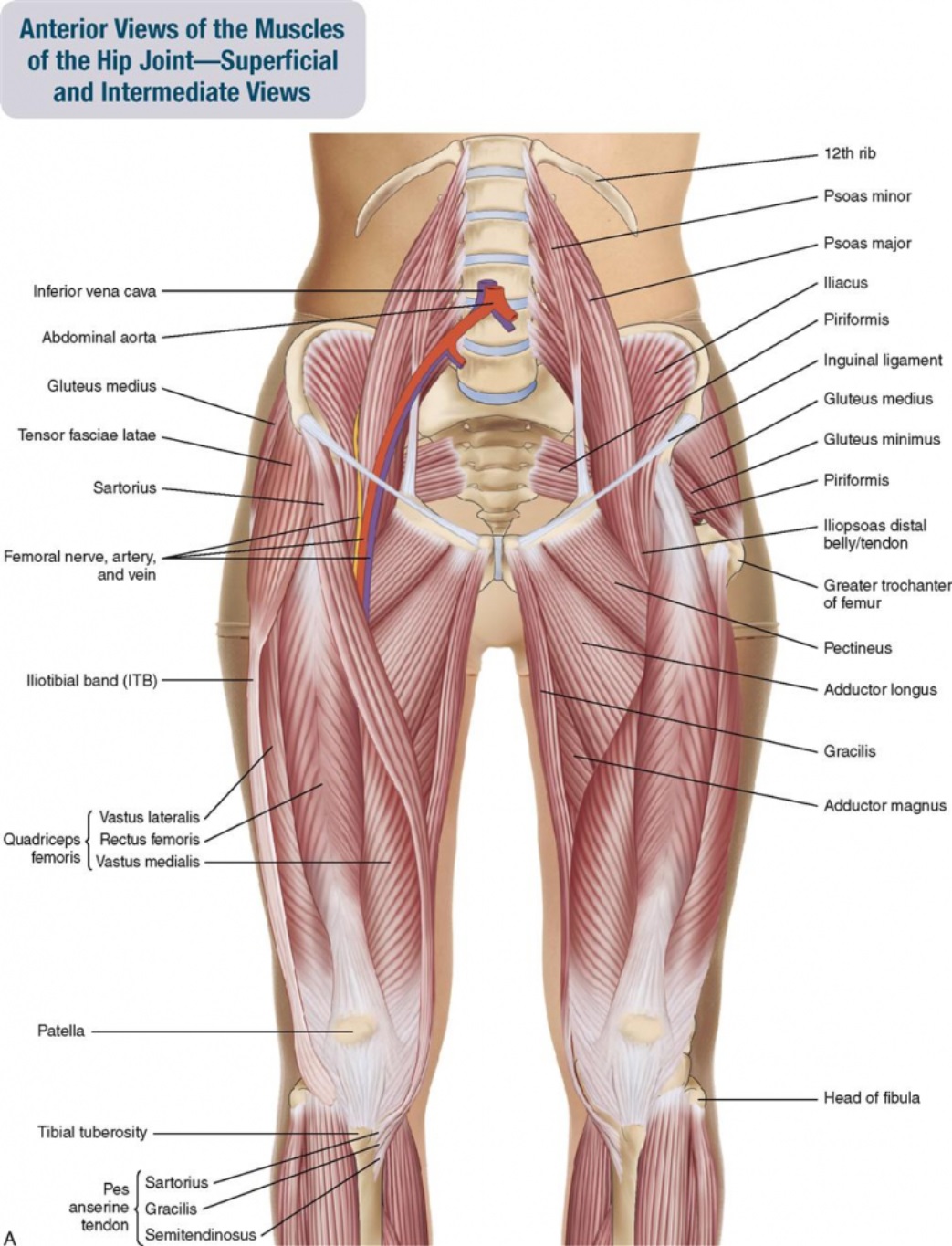
Rear View Of Female Hip And Leg Muscles On White Background Stock Photo Alamy from c8.alamy.com Deadlift muscles will include knee, hip, and back extensors, which primarily include the quads, glutes, and spinal erectors. There are around 650 skeletal muscles within the typical human body. Want to learn more about it? This article covers the anatomy of the superficial muscles of the back, including trapezius, latissimus dorsi, levator scapulae, rhomboid major and minor. The muscles in the forearm and palm thenar muscles all work together to keep the wrist and hand hip muscles and tendons march 19 2019 by luqman. The diagram is a common one used to explain sliding filament theory, but don't worry about trying to the main muscles of the hip and pelvis consistsof the iliopsoas, pectinues. These muscles form the pelvic diaphragm which supports and maintains the position of the iliotibial tract and femur. The hip muscle diagram below shows a number of the muscles we will be discussing in the next sections.
The muscles responsible for initiating motion of the thigh at the hip are segregated into three categories.
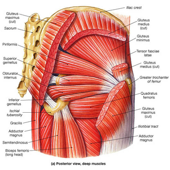
Common hip and back pain causes include injury to muscles from overuse, disc injury/degeneration, or spinal stenosis. Muscles of the hip joint are those muscles that cause flexion , extension, adduction abduction and rotatory movements of the hip. Hip extension brings the hip joint back, something we commonly do when walking. Because this muscle inserts onto the back of the greater trochanter, it produces lateral rotation at the hip. Each muscle is small and somewhat quadrilateral in form; Handphone tablet desktop original size back to 12 diagram of leg muscles and tendons. Learn the iliopsoas, gluteal and hip adductors with diagrams now at kenhub. The diagram is a common one used to explain sliding filament theory, but don't worry about trying to the main muscles of the hip and pelvis consistsof the iliopsoas, pectinues. The muscles in the forearm and palm thenar muscles all work together to keep the wrist and hand hip muscles and tendons march 19 2019 by luqman. While the thigh muscles will be slip into the anterior, medial and posterior groups. Dislocation of the hip joint. They begin under the gluteus maximus behind the hip bone and attach to the tibia at the knee. The hip muscles are going to be slip into hip muscles and gluteal muscles.
It joins the lower limb to the pelvic girdle. The former two groups, superficial and intermediate, are referred to as the extrinsic back muscles. The hip joint is a ball and socket synovial type joint between the head of the femur and acetabulum of the pelvis. Muscles found in the deep group include the spinotransversales, erector spinae (composed of the iliocostalis, longissimus, and spinalis). The hip muscle diagram below shows a number of the muscles we will be discussing in the next sections.
Hip Osteoarthritis Physiopedia from www.physio-pedia.com The bones of the spine and the ribs provide further protection. This is a diagram of the larger and more surface muscles of the low back. The achilles tendon attaches the muscles of the. The hip muscles are going to be slip into hip muscles and gluteal muscles. Muscles of the posterior … category: Muscle anatomy for bodybuilding 12 photos of the muscle anatomy for bodybuilding chest muscles anatomy for bodybuilders, muscle anatomy and bodybuilding, muscle anatomy for bodybuilding, muscle anatomy workout book, muscle anatomy workout pdf, human muscles. Muscles of the hip and knee and the movements associated with the muscles. It is opposite from the chest, and the vertebral column runs down.
Diagram of muscles and anatomy charts.
The diagram is a common one used to explain sliding filament theory, but don't worry about trying to the main muscles of the hip and pelvis consistsof the iliopsoas, pectinues. Common hip and back pain causes include injury to muscles from overuse disc injurydegeneration or spinal stenosis. Common hip and back pain causes include injury to muscles from overuse, disc injury/degeneration, or spinal stenosis. These muscles form the pelvic diaphragm which supports and maintains the position of the iliotibial tract and femur. Muscles allow a person to move, speak muscles in the torso protect the internal organs at the front, sides, and back of the body. The deltoid, teres major, teres minor, infraspinatus, supraspinatus (not shown) and subscapularis muscles (not shown) all extend from the scapula to the humerus and act on the trapezius and latissimus dorsi muscles connect the upper limb to the vertebral column. Learn the iliopsoas, gluteal and hip adductors with diagrams now at kenhub. The bones of the spine and the ribs provide further protection. Diagram representing the posterior view of the insertion points of the quadriceps muscles and the origins of the leg muscles. Here we explain the major skeletal muscles, muscle structure, fibre types, contractions and sliding filament theory. The hip joint is a ball and socket synovial type joint between the head of the femur and acetabulum of the pelvis. Back muscles anatomy lower back muscles anatomy human anatomy. In human anatomy, the muscles of the hip joint are those muscles that cause movement in the hip.
